
FIGURE 1.
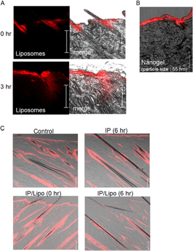
Effects of IP with cationic liposomes on physiological conditions of rat dorsal skin. A, time-dependent change in the distribution of rhodamine-labeled cationic liposomes (red) in the skin after IP treatment. B, IP-induced epidermal penetration of cationic nanogel labeled with rhodamine (red). C, phalloidin immunohistochemical analysis of F-actin (red) in the skin. Control means the intact skin without IP/liposome treatment. IP (6 h) means the skin at 6 h after IP without liposomes. IP/Lipo (0 h) and IP/Lipo (6 h) are the skins at 0 and 6 h after IP with liposomes. Bar indicates 100 μm.