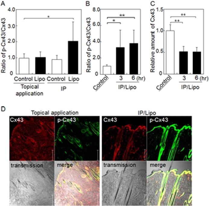
FIGURE 3.

IP with cationic liposomes induces Cx43 phosphorylation in rat skin. Western blot analysis of the relative amount of phosphorylated Cx43 compared with total Cx43 in skin treated with liposomes followed by additional incubation for 0 h (A), and 3 or 6 h (B). C, Western blot analysis of the relative amount of total Cx43 in skin treated with liposomes followed by additional incubation for 3 or 6 h. D, immunohistochemical analysis of Cx43 (red) and phosphorylated Cx43 (green) in skin treated with IP with liposomes. The data are represented as the average with standard deviation (n > 3). **, p < 0.01, *, p < 0.05. Bar indicates 100 μm.
