
FIGURE 8.
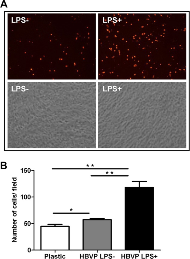
PBL adhesion to HBVPs treated with LPS. A, fluorescently labeled PBLs were added onto a confluent monolayer of LPS-treated HBVPs and washed after 2 h. Original magnification, ×200. B, quantification of adhered cells. Values correspond to the arithmetic mean ± S.D. of three different fields belonging to a representative experiment. *, p < 0.05; **, p < 0.005. Error bars represent S.D.