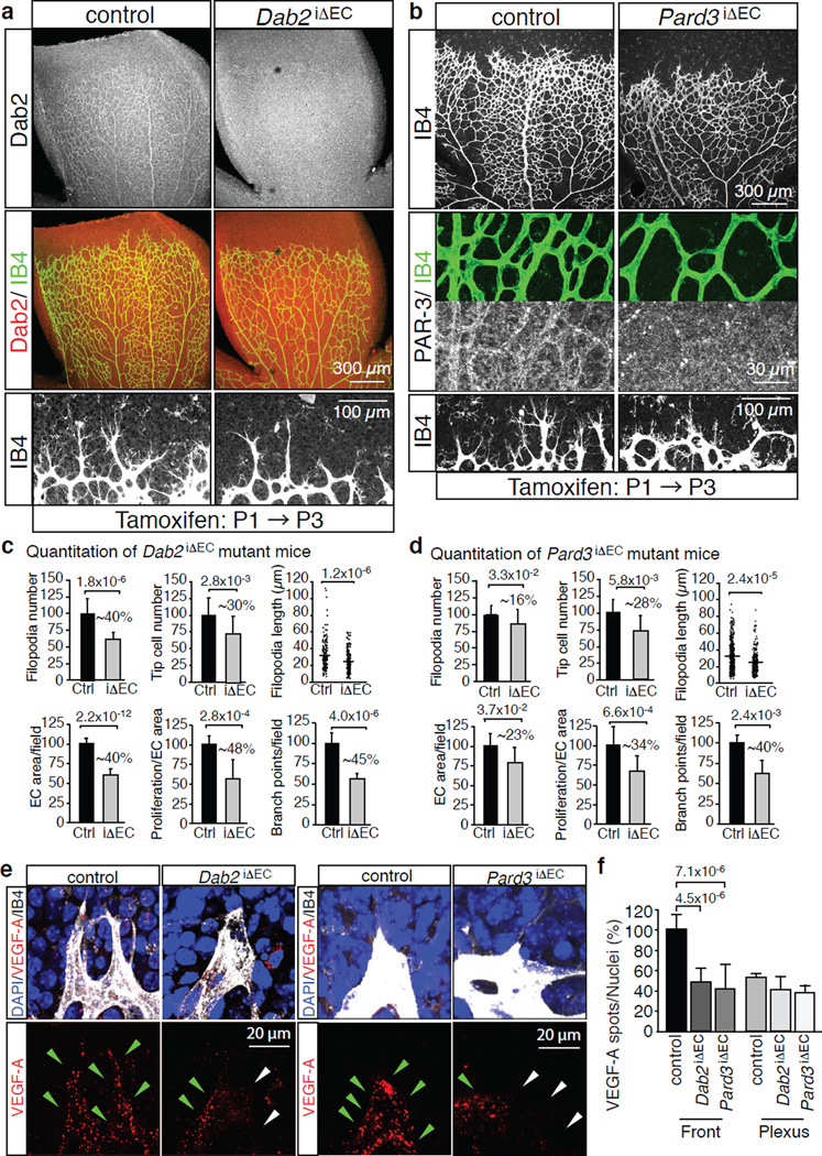
Figure 5. Endothelial Dab2 and PAR-3 regulate angiogenic vessel growth.
a, Overview of the P6 control and Dab2iΔEC retinal vasculature. Anti-Dab2 (white/red) staining is shown in top panels. ECs, Isolectin B4 (IB4). Bottom panels show higher magnification of the angiogenic front.
b, Defects in the P6 Pard3iΔEC retinal vasculature. Anti-PAR-3 and IB4 (green) staining in middle panels show successful deletion of PAR-3 in Pard3iΔEC vessels. Residual round signals correspond to autofluorescent blood cells. Bottom panels show higher magnification of the angiogenic front.
c, d, Quantitation of filopodia number and length, tip cell number, EC-covered area, EC proliferation and vessels branch points in Dab2 mutant (iΔEC) (c) or Pard3 mutant (d) retinas compared to the corresponding control littermates (Ctrl). Dab2 mutant n=7, Pard3 mutant n=3. Percentage of reduction is indicated. Data represent the means±s.d. P values, two-tailed Student’s t-test.
e, Reduced uptake of labelled VEGF-A (red) at the Dab2iΔEC or Pard3iΔEC angiogenic front compared to control littermates. Green arrowheads indicate VEGF-A spots, white arrowheads marks ECs with no or little uptake.
f, Statistical analysis of internalised Alexa-coupled VEGF-A in the angiogenic front and central plexus. Data represent the means±s.d. of 6 independent experiments. P values, two-tailed Student’s t-test.

