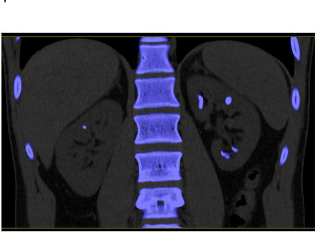
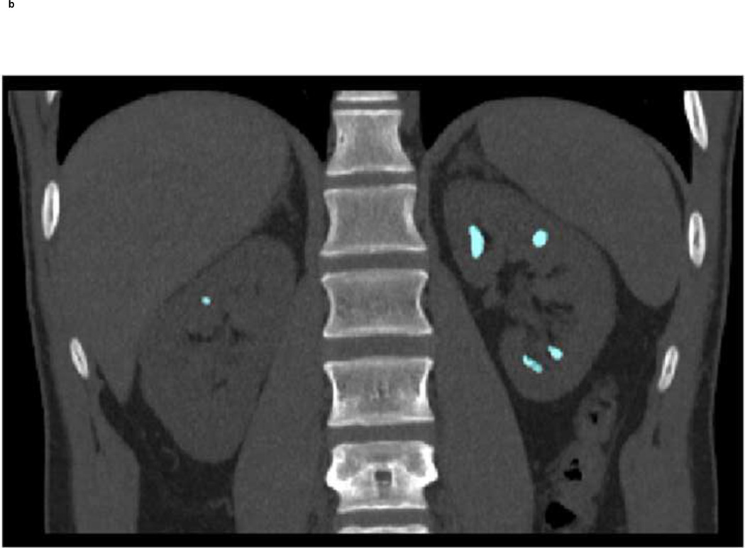
Fig. 7.
A clinical example demonstrating the feasibility of using dual-source DECT with tin filtration to differentiate non-UA stone types. Using commercially software (“Kidney Stone”, Syngo CT Workplace, Siemens Healthcare), the stones were coded in blue (a) indicating a non-UA stone type. With dual-source DECT and additional tin filtration on the high energy tube, it was possible to characterize these stones into Group-4, which was defined to include three stone types: calcium oxalate mono- or dihydrate or brushite. Group-4 is coded in turquoise (b). The stones were subsequently removed from the patient with percutaneous nephrolithotomy and were confirmed to be pure calcium oxalate dihydrate by both Fourier transform infrared spectroscopy and micro-CT.


