Figure 1.
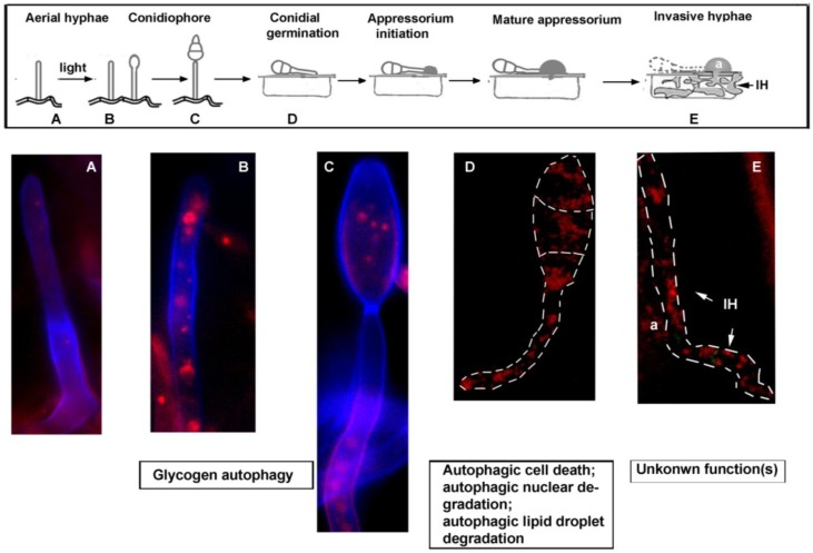
Schematic diagram of M. oryzae pathogenic life cycle, and natural induction of autophagy. Schematic representation of the pathogenic life cycle of M. oryzae (boxed), with corresponding steps assessed for autophagy (RFP-Atg8) induction depicted in (A–E). Basal level of RFP-Atg8 is undetectable in the aerial hyphae grown in the dark (A). Upon photo-induction, RFP-Atg8 is naturally induced in the aerial hyphae (B), as well as in the conidiophore (C). For (A)–(C), Magnaporthe strain expressing RFP-Atg8 was grown on PA (prune agar) medium, co-stained with Calcofluor White and analysed by confocal microscopy. RFP-Atg8 was also naturally induced during conidial germination (D) and in invasive hypha (E). For (D)–(E), dashed lines were used to delineate the outline of the analyzed fungal structures. a, appressorium; IH, invasive hypha. Arrows in (E) mark primary invasive hypha (36–40 hpi (hours post inoculation)).

