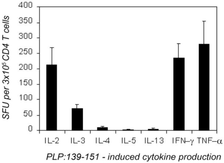
Figure 1.
Overall type-1 cytokine profile of proteolipid protein (PLP):139-151-specific CD4 cells 7 days after the immunization with PLP:139-151. CD4 cells (3 × 10⁵/well) purified from draining lymph nodes (dLN) were tested with naive syngeneic (SJL) LN cells (5 × 10⁵/well) in the presence of a maximally stimulatory dose of PLP:139-151 peptide (20 mM). Enzyme-linked immunospots (ELISPOTs) (SFU) were counted by an image analyzer. The results were obtained in four independent experiments, each with four–six mice tested individually. These data are expressed as the mean + SD spots forming cells per 3 × 10⁵ CD4 cells tested.

