Figure 3.
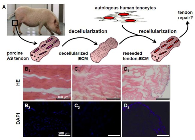
Dezellularization and recellularization of porcine Achilles tendons for tendon repair.Schematic diagram of the decellularization of porcine Achilles (AS) tendon as a scaffold for reseeding with autologous human hamstring tenocytes to use the resulting constructs for tendon defect coverage (A). HE and 4',6-diamidino-2-phenylindole (DAPI) staining of a native (B1, B2), decellularized (C1, C2) and a porcine Achilles (AS) tendon ECM recolonized with human hamstring tenocytes (D1, D2). Clefts arise between collagen fiber bundles in response to decellularization. Scale bars: 100 µm (B1, C1, D1), 200 μm (B2, C2, D2).

