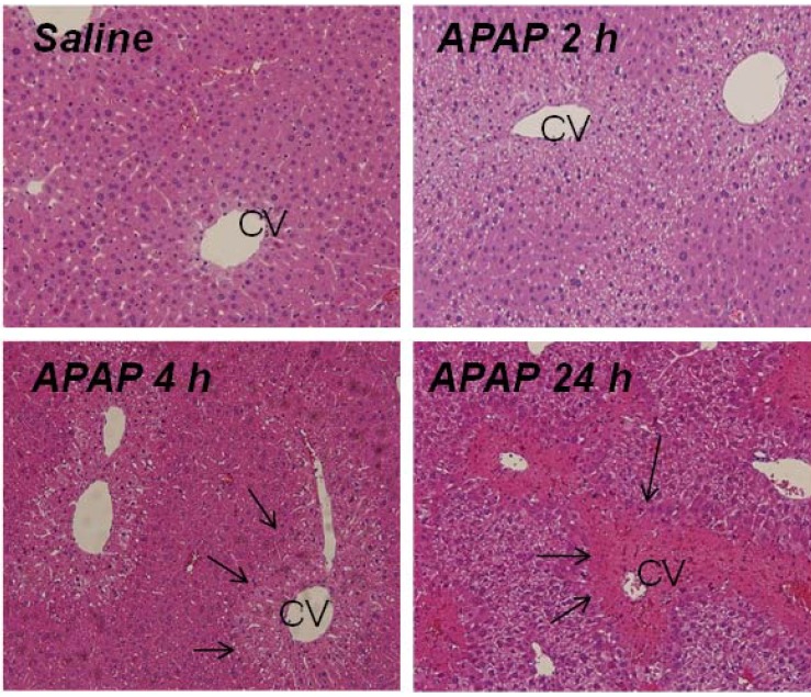
Figure 2.
H and E stained liver sections of representative B6C3F1 male mice treated with APAP (200 mg/kg IP). By 2 h, centrilobular pallor was apparent in the cells surrounding the central vein (CV). By 4 h, frank necrosis (demonstrated by arrows) was present in the region surrounding the CV, which extended to bridging necrosis at 24 h post-APAP.

