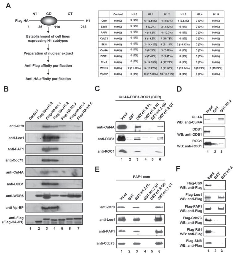
Figure 1. Identification of the Cul4A and PAF1 complexes as interaction partners of linker histone H1.2.
(A) The six human H1 subtypes and their associated factors were isolated from nuclear extracts of HeLa S3 cells stably expressing Flag-HA-H1 subtypes through sequential immunoaffinity chromatographies. Three independent affinity purifications from HeLa nuclear extracts expressing Flag-HA-H1 subtypes were used for the MudPIT mass spectrometry assay. Among the reproducible and significant (p-value < 0.001) proteins identified in all three analyses, all subunits of both Cul4A and PAF1 complexes were detected by multiple peptides. The table summarizes the peptide count and the amino acid coverage of Cul4A and PAF1 components co-purified with H1 subtypes.
(B) The purified samples shown in (A) were resolved on 4–20% SDS-PAGE, and the presence of the Cul4A and PAF1 complexes was confirmed by immunoblot analysis
(C) The reconstituted Cul4A-DDB1-ROC1 (CDR) E3 ligase complex was incubated with glutathione-Sepharose beads containing GST alone, GST-H1.2 full length (FL), GST-H1.2 N-terminal tail (NT), GST-H1.2 globular domain (GD) and GST-H1.2 C-terminal tail (CT). The bound proteins were analyzed by immunoblotting with the indicated antibodies. Input corresponds to 10% of the CDR complex used in the binding reactions.
(D) GST alone (lane 2) or GST-H1.2 (lane 3), immobilized on glutathione-Sepharose beads, was incubated with recombinant Cul4A, DDB1 and ROC1. After washing with washing buffer, the bound proteins were immunoblotted with anti-Flag antibody. Ten percent of the input proteins were examined by immunoblotting (lane 1).
(E) For the pull-down experiments, the purified PAF1 complex was incubated with GST or the indicated GST-H1.2 fusions and subjected to immunoblotting after extensive washing. Input lane represents 10% of the PAF1 complex used in the binding reactions.
(F) GST pull-down assays were conducted as described in (D), but using Flag-tagged subunits of the PAF1 complex that were individually expressed and purified from Sf9 cells. Binding of each protein was analyzed by immunoblotting. Lane 1 represents 10% of the input. See also Figure S1.

