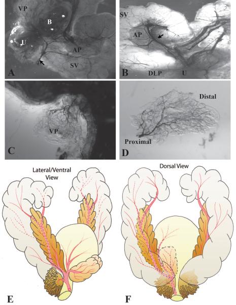
Figure 1.
Visualization of the prostatic vascular anatomy by intracardiac perfusion of India ink. A. Low magnification brightfield image of the ventral and lateral side of adult male mouse urogenital tract. The blood vessels were filled with black India ink. Black arrow indicates the main artery supplying the male urogenital tract. Note that left lobe of the ventral prostate was removed to visualize the vessels near the neck of the bladder. B. Low magnification brightfield image of the dorsal side of adult mouse male urogenital tract. Black arrow indicates the main artery supplying the seminal vesicle and anterior prostate. C–D. Images of ventral prostate showing that the vessels supplying the lobe enter close to the urethra and arborize distally. U Urethra. VP Ventral prostate. DLP Dorsalateral prostate. AP Anterior prostate. SV Seminal vesicle. B Bladder. E–F. Schematic illustrations of the vascular anatomy of the adult male mouse urogenital tract. E. A lateral and ventral view. F. A dorsal view. The main artery on the ventral aspect of the bladder runs laterally onto each side of the male urethra and branches into three vessels near the neck of the bladder: the ventral branch supplies the bladder and the ventral prostate; the central branch supplies the seminal vesicle and the anterior prostate; the dorsal branch supplies the dorsolateral prostate.

