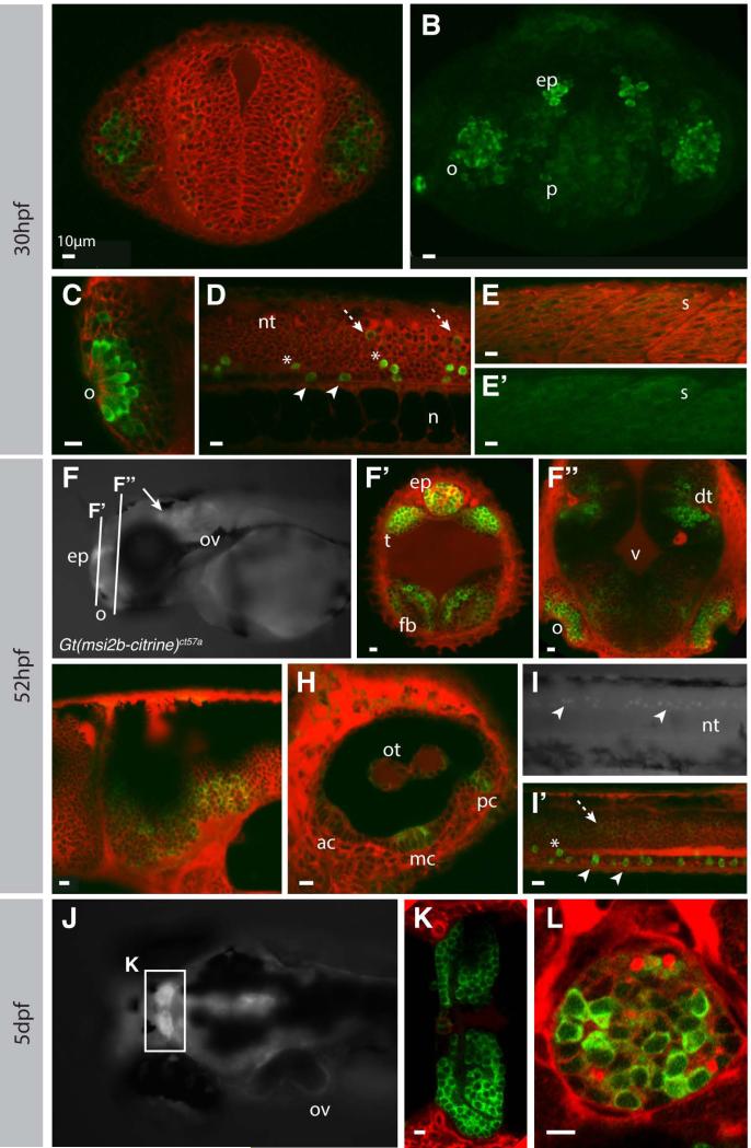
Figure 4. Msi2b is expressed in distinct neuronal populations in zebrafish.
Confocal imaging of live gt(msi2b-citrine)ct57a embryos depicts cytoplasmic expression of Msi2b in zebrafish embryos (green) at 30 hpf (A-E’), 52 hpf (F-I’) and 5 dpf (J-L). Counterstaining by BODIPYTR (red). Olfactory region, o; pallium, p; epiphysis, ep; otic vesicle, ov; neural tube, nt; notochord, n; somite, s; telencephalon, t; forebrain, fb; dorsal telencephalon, dt; midbrain-hindbrain boundary, mhb; otoliths, ot; anterior, median and posterior cristae are respectively ac, mc and pc. Scale bars: 10 μm. (A, B) Frontal view. Single plane imaging (A) and respective confocal projection (B) of Msi2b-Citrine expression in the olfactory placode and pallium, with lower levels of expression in the forebrain. (C-E’) Lateral views. (C) Msi2b is highly expressed in the olfactory placode. (D) Expression of Msi2b in three distinct subpopulations of neurons in the ventral neural tube highly expressing cells within (white arrowhead) and dorsally (asterisks) to the floor plate, and low expression in the periphery of the neural tube (dashed white arrow). (E, E’) Low levels of Mis2b are detected in the somites. (F, G-I’) Lateral views. (F’,G”) Coronal sections. (F) Msi2b is expressed in the epiphysis, olfactory organs, otic vesicles and CNS (white arrow). (F’, F”) Frontal views, optical sections. Expression in the dorsal thalamus and epiphysis as well as in the forebrain and olfactory organs. (G) Msi2b is expressed in the ventral cells of the hindbrain. (H) Expression in the otic vesicle is localized to cells of the otoliths and in the central cells of the cristae of the semicircular canals. (I, I’) Expression of Msi2b in the ventral cells of the neural tube (white arrowhead). Cells dorsal to the floor plate (asterisks) and low expression in the periphery of the neural tube (dashed white arrow). (J) Dorsal view of zebrafish head at 5dpf. Msi2b is expressed in the habenula (K) and is restricted to a subset of neurons in the epiphysis (L).

