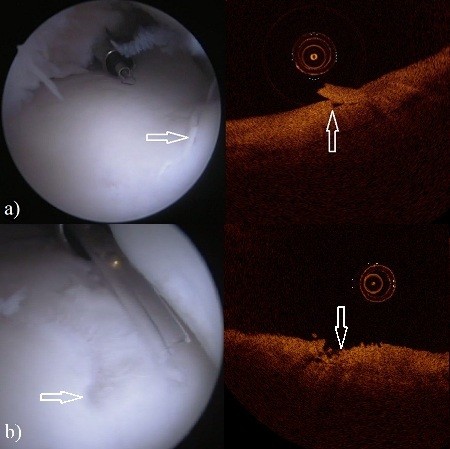
Figure 2.

The arthroscopy (left) and OCT (right) images of the third carpal bone in the intercarpal joint. a) Lesion (arrows) in the arthroscopic image (left) and in the OCT image (right) with ICRS score of 2. b) Lesion (arrows) in the arthroscopic image (left) and in the OCT image (right) with ICRS score of 3.
