
Figure 1.
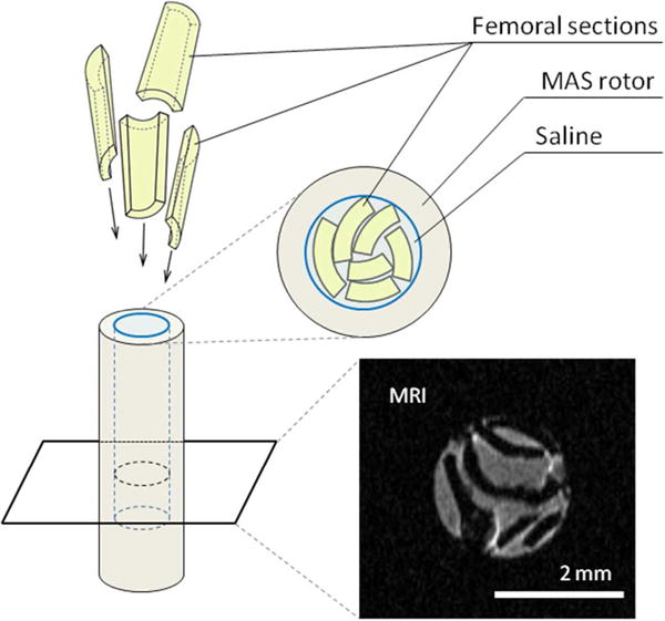
Schematic representation of the packing of femoral mid-diaphyseal sections into the NMR rotor. A representative 1H MRI image of a transverse cross-section of the rotor shows the actual arrangement of the bone sections (black) surrounded by saline (grey). Images confirm that the bone fragments remained intact. No powdering, chemical treatment or drying was required to probe the molecular structure of the mouse femora.