Figure 3.

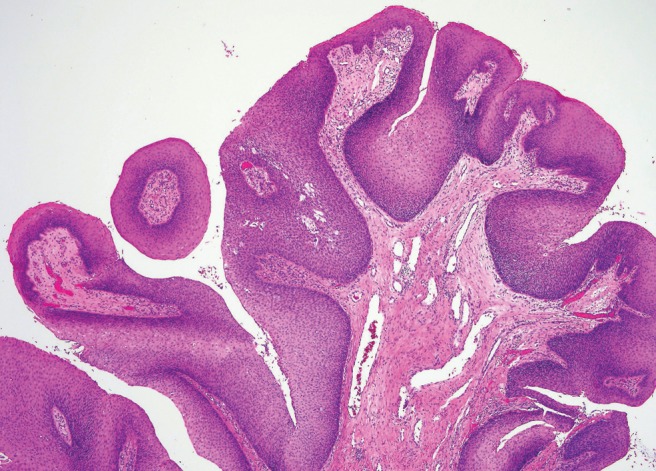
Low-power magnification, hematoxylin and eosin stain depicting exophytic papilloma with branching, exophytic proliferations, and a fibrovascular core, lined by well-differentiated stratified squamous epithelium.

Low-power magnification, hematoxylin and eosin stain depicting exophytic papilloma with branching, exophytic proliferations, and a fibrovascular core, lined by well-differentiated stratified squamous epithelium.