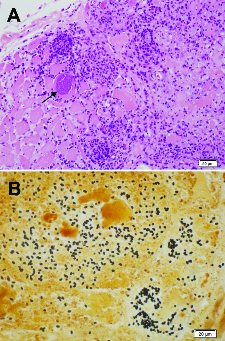
Figure 1.
Light micrographs of muscle biopsy tissue from a 67-year-old man (case-patient A), New South Wales, Australia, showing microsporidial myositis caused by Anncaliia algerae. A) Necrotizing myositis with prominent inflammation and spores within the necrotic cytoplasm of a myocyte (arrow). Hematoxylin and eosin stain. Scale bar indicates 50 μm. B) Numerous dark brown to black, 3- to 4-μm ovoid spores in necrotic myocytes. Warthin-Starry stain. Scale bar indicates 20 μm.

