
Figure.
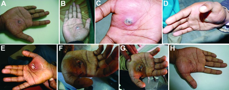
Progression of lesion on palm of researcher caused by buffalopox virus infection, India. A) Small vesicle on postinjury day 5. B) Pustule with a central area of necrosis on postinjury day 7. C) Pustule with edema on postinjury day 9. D) Increase in edema on postinjury day 10 (cyanosis is not apparent). E) Lesion after surgical excision on postinjury day 11. F) Lesion on postinjury day 19 (healing was erratic). G) Blackening and thickening around surgical site on postinjury day 30, which then extended to a wider circumference. H) Healed lesion with a black eschar on postinjury day 85.