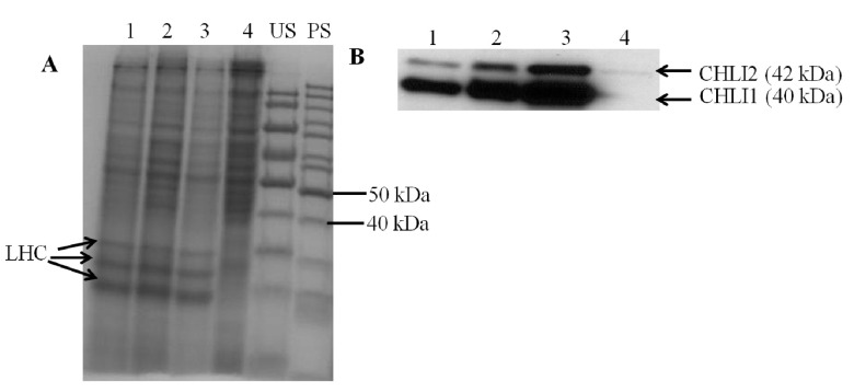
Figure 11. SDS-PAGE and Western analyses.
( A) A stained protein gel. Lanes 1, 2, 3 and 4 represent chli1-8, chli1-7, 4A+ and 5A7, respectively. Light harvesting complex (LHC) protein bands are labeled. PS and US denote pre stained and unstained molecular weight protein ladders, respectively. Total cell extract of different strains were loaded on equal Chl basis (4 µg of Chl) in lanes 1, 2 and 3. In lane 4, 40 µg of protein (the maximum amount of protein that can be loaded on a mini protein gel) was loaded as 5A7 lacks Chl. ( B) Western analyses using a CHLI1 antibody generated against the Arabidopsis CHLI1 protein. Lanes 1, 2, 3 and 4 represent chli1-8, chli1-7, 4A+ and 5A7, respectively. CHLI1 (40 kDa) and CHLI2 (42 kDa) proteins detected by the antibody are labeled.

