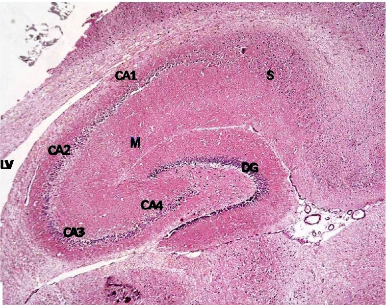
Figure 6. Section from non-diabetic control showing the different areas of the hippocampal formation where the hippocampus proper is formed of the Cornu Ammonis (CA) as CA1, CA2, CA3 & CA4 regions, and is continued as subiculum (S).
Dentate gyrus (DG) is seen surrounding CA4 by its upper & lower limbs. Note lateral ventricle (LV) related to CA1 & CA2. M denotes molecular layer inside concavity of CA and of DG. (H & E ×40).

