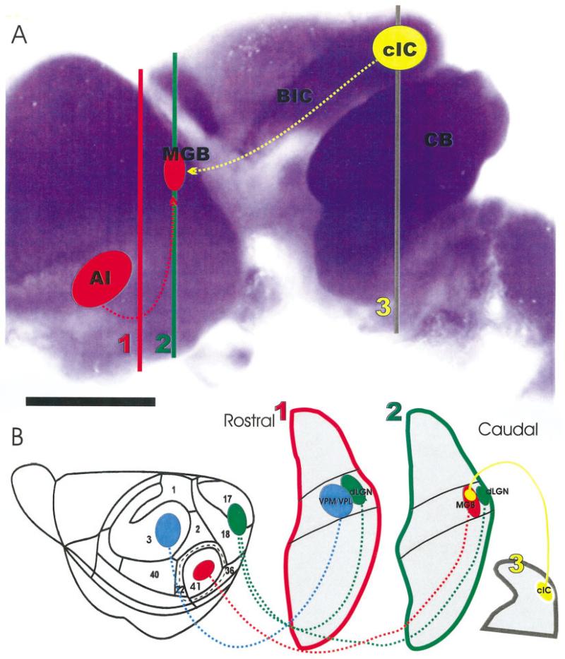
Fig. 1.
The organization of NeuroD expression in the brain, the distribution of injection sites into the presumed auditory cortex and inferior colliculus, and the nuclear and connections organization as revealed in coronal sections is shown. A: Note that the cerebellum, parts of cortex, and the inferior colliculus show strong β-galactosidase reactivity, indicating up-regulation of NeuroD in these areas. The position of the presumptive auditory cortex (AI), medial geniculate body (MGB), central nucleus of the inferior colliculus (cIC) is shown in this lateral view of an embryonic day 18 brain. B: Other injection sites in the somatosensory cortex (blue) and the visual cortex (green) as well as their corresponding thalamic relay nuclei (ventrolateral/ventromedial posterior nucleus, VPM/VPL; dorsal lateral geniculate nucleus, dLGN) are shown at the approximate section levels as indicated in A. BIC, brachium of the inferior colliculus; CB, cerebellum. Scale bar = 1 mm in A.

