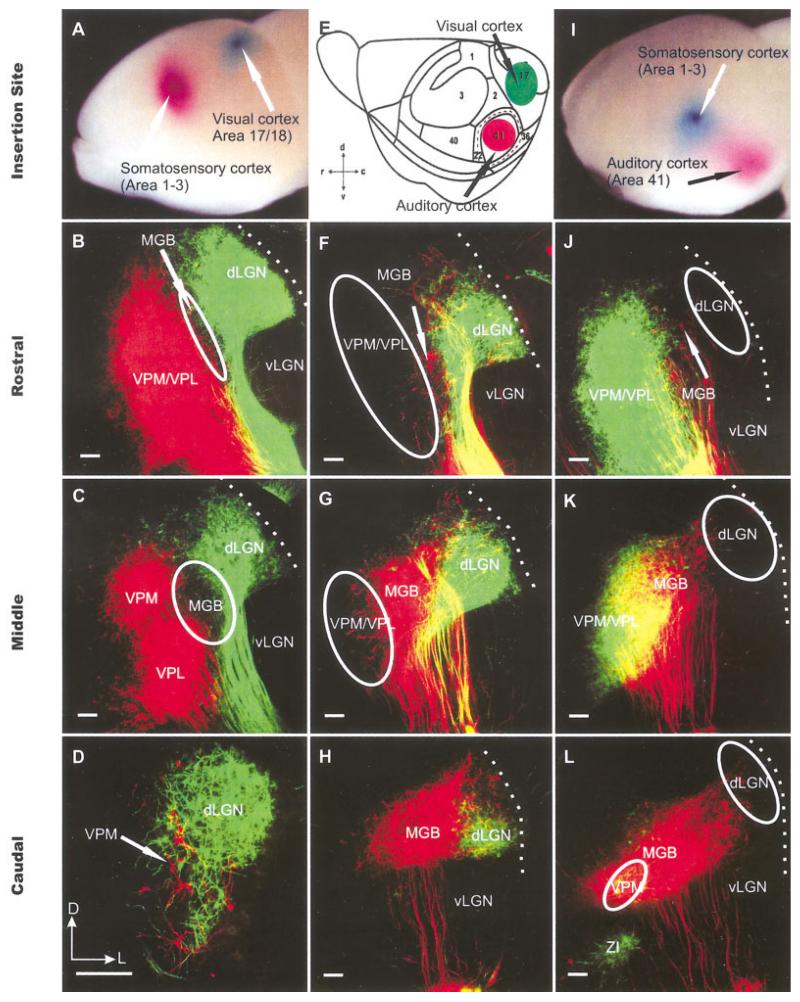
Fig. 2.
The organization of thalamocortical projection neurons as revealed in coronal sections after double dye insertions at embryonic day (E) 18.5. A,E,I: Specific sets of thalamic neurons were double labeled from auditory, visual, and somatosensory cortex with the spectrally distinct lipophilic dyes PTIR 271 (colored in green) and PTIR 278 (colored in red). This strategy allowed us to distinguish between caudomedial groups labeled from the presumptive auditory cortex, a lateral group labeled from the presumptive visual cortex, and rostromedial groups labeled from the presumptive somatosensory cortex. F-H,J-L: In agreement with the known distribution of these thalamic nuclei in adult mice, we find a caudal group labeled after presumptive auditory cortex insertions (red). F-H: More rostrally, this presumptive MGB population is dorsolateral capped by a population that is consistently labeled after presumptive visual cortex insertions. B-D,J-L: Even more rostral, we find a population only labeled after presumptive somatosensory cortex insertions, which we refer to as VPM/VPL. We can distinguish, therefore, between the different corticothalamic projections nuclei as early as E18.5. E: The cortical map follows that of Stiebler et al. (1997). dLGN; dorsal lateral geniculate nucleus; MGB, medial geniculate body; vLGN, ventral lateral geniculate nucleus; VPL, ventrolateral posterior nucleus; VPM, ventromedial posterior nucleus; ZI, zona incerta. Dotted lines indicate thalamic surface. D,E: Orientation is indicated for these and all other coronal sections of Figures 2-9: d, dorsal; c, caudal; v, ventral; r, rostral; D, dorsal; L,, lateral. Scale bars = 100 μm in B-D, F-H,J-L.

