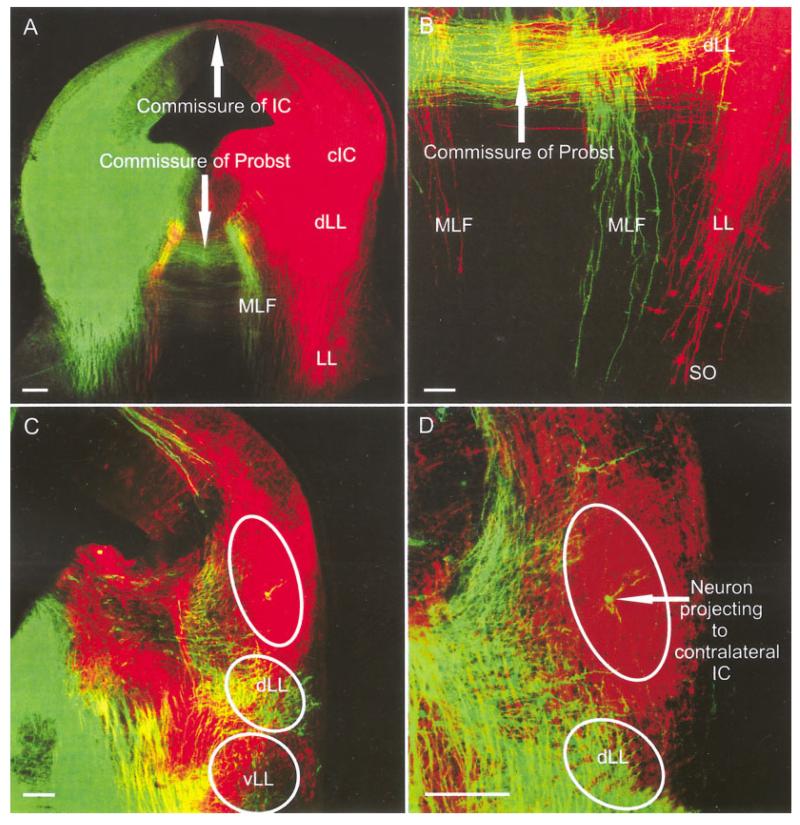
Fig. 4.
IC insertions and labeled fibers and neurons in the brainstem at embryonic day 14.5. A,B: Insertions into the IC-labeled ipsilateral LL fibers that extended toward to the SO region where few neurons were retrogradely filled. We also found that LL fibers crossed the commissure of Probst to reach the contralateral LL. A-D: These fibers did not only extend to dorsal LL (dLL) neurons (B-D) but also projected along the medial longitudinal fascicle toward the contralateral brainstem reticular formation (A,B). A,C,D: In addition, contralateral central nucleus of the IC (cIC) fibers crossed to invade the cIC as well as filling their cells of origin. D: These contralateral cIC neurons were most frequently found dorsal in central nucleus of the IC. B: We could not label any fibers projecting beyond the superior olive, which showed only few neurons projecting to the ipsilateral lateral lemniscus. IC, inferior colliculus; LL, lateral lemniscus; MLF, medial longitudinal fascicle; SO, superior olive. Scale bars = 40 μ in A; 10 μm in B,C; 20 μ in D.

