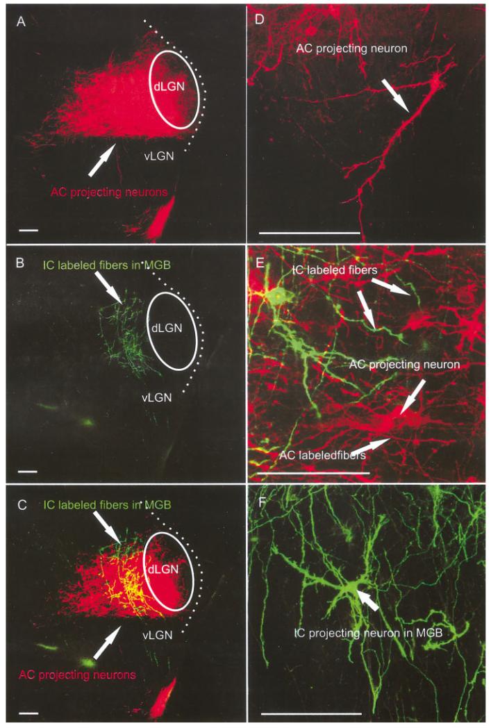
Fig. 7.
AC and IC insertions reveal more extensive cell and fiber overlap at E18.5. A-C: Caudal IC insertions labeled fibers overlapping with MGB neurons labeled after auditory cortex insertions. D-F: Higher magnifications showed the more elaborate dendritic trees of the MGB neurons (D), IC projecting neurons (F), and a prominent overlap of dendrites and axons of retrogradely filled neurons as well as anterogradely filled fibers (E). AC, auditory cortex; IC, inferior colliculus; dLGN; dorsal lateral geniculate nucleus; MGB, medial geniculate body; vLGN, ventral lateral geniculate nucleus. Scale bars = 10 μ in A-C; 60 μ in D-F.

