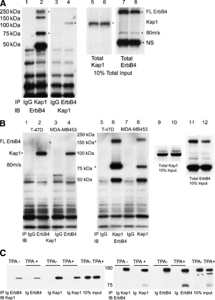
FIGURE 2.
Reciprocal coimmunoprecipitation of ErbB4 and Kap1 in breast cancer cell lines. A, coimmunoprecipitation from NRG-treated BT-474 cells. Five hundred micrograms of BT-474 cell lysate were immunoprecipitated with 2 µg of total rabbit IgG, anti-Kap1, or anti-ErbB4. Immune complexes were isolated using protein A/G beads and analyzed on 4% to 12% NuPAGE Bis-Tris gels. Portions of the lysates were analyzed directly in lanes 5 to 8. Immunoblots were probed with the reciprocal antibodies to those used for immunoprecipitation. Asterisks mark the full-length and 80-kDa cleaved forms of ErbB4 (lanes 2, 7, and 8) or Kap1 (lanes 4–6). NS, nonspecific band on ErbB4 blots. B, coimmunoprecipitation of ErbB4 and Kap1 from NRG-treated T-47D and MDA-MB453 cells. Portions of the lysates were analyzed directly without immunoprecipitation in lanes 9 to 12. C, HEK293T cells were transfected with 1 µg each of the plasmids encoding full-length ErbB4 and Flag-Kap1. After 24 h, the cells were switched to starvation medium (0.1% FBS in DMEM) for 16 h and then mock stimulated with DMSO vehicle (TPA−) or with 100 ng/mL TPA (TPA+) in DMSO. Lysates were immunoprecipitated with anti-Kap1, anti-ErbB4, or control IgG (Ig). Kap1 and ErbB4 were identified by immunoblotting, with ~100-kDa Kap1 and full-length and cleaved ErbB4 forms. Blotting for GAPDH (not shown) verified even loads for 10% input tracks. Data represent at least two biological replicates in identical format plus two similar biological replicates (A and B).

