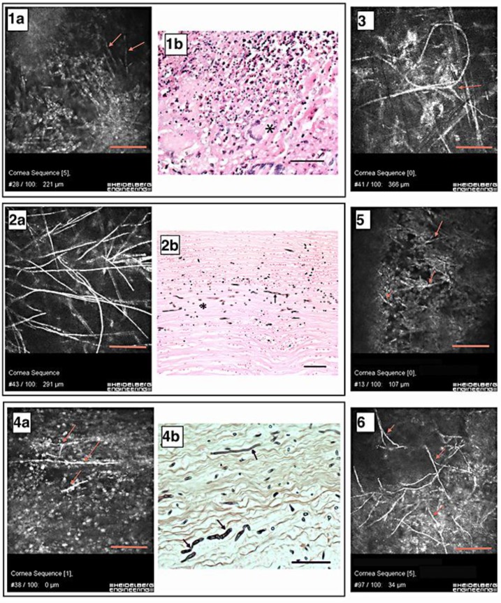
Fig. 2.
IVCM results and histopathological images. Numbers correspond to patient numbers in table 1. Scale bars = 100 μm. 1a Hyphae in the corneal stroma (red arrows). 1b Corneal button with severe granulomatous inflammation. Asterisk: foreign body giant cell. HE. 2a A stromal meshwork of hyphae. 2b Cornea with septated hyphae (arrow) and cross-sectioned hyphae (asterisk). Grocott. 3 Interlocking and irregularly shaped hyphae with a bifurcature (red arrow). 4a Red arrows show isolated hyphae within the epithelium. 4b Cornea with hyphae of various dimensions (arrows). Grocott. 5 Red arrows show thick bifurcating hyphae. 6 Red arrows show epithelial hyphae.

