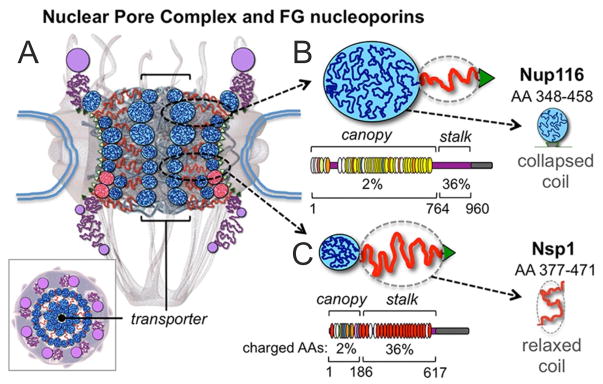
Figure 1.

Diagrams of the NPC and its disordered FG nups. (A) Cross-section featuring the NPC ring-scaffold (center), cytoplasmic fibers (top), and nuclear basket (bottom). Intrinsically-disordered FG nups are outlined in blue (collapsed coils) and red (extended coils) lines forming the transporter/plug gate structure in the NPC channel [11]. A top view is also shown (bottom left). (B) The S. cerevisiae FG nups Nup116. As a representative of ‘cohesive’ nups it primarily adopts collapsed coil/’canopy’ configurations (blue line, globule). Below FG repeats are shown as thin ovals (GLFG in yellow; FxFG in red) and the amount of charged AA is indicated. The portion of Nup116’s canopy (AA 348–458) is highlighted. (C) Nsp1 as a representative of ‘repulsive’ nups. It adopts a relaxed-to-extended ‘stalk’ region (red line) and it has a high content of charged AAs and features mainly FxFG motifs (bottom). The portion of Nsp1’s stalk (AA 377–471) is highlighted.
