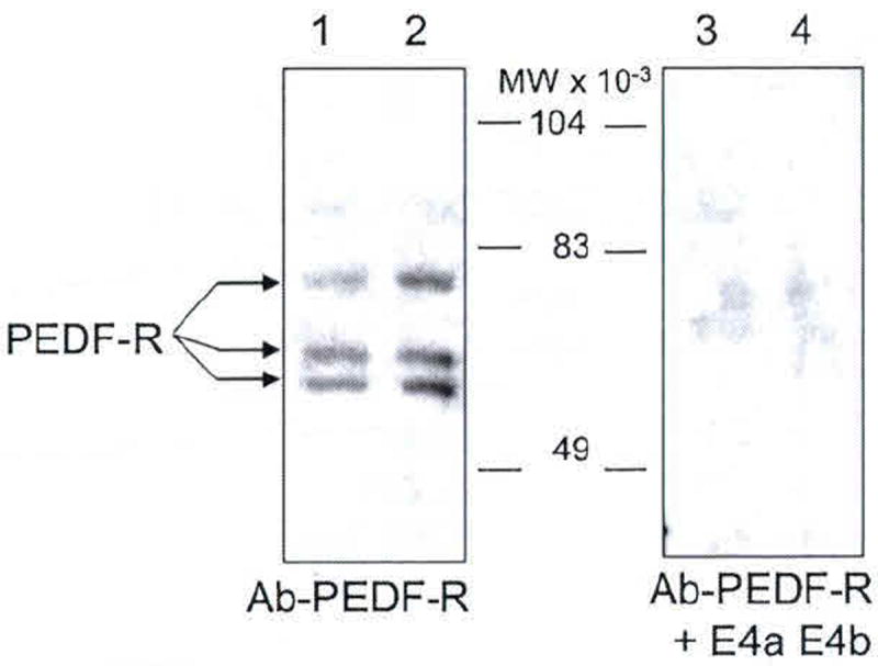
Fig. 102.4.

Western blot of native PEDF-R from retina R28 cells. Membrane fractions obtained from R28 cells were resolved by SDS-PAGE. Total protein loaded in lanes 1–4 was 6 μg each. Lanes 1 and 2, and lanes 3 and 4 were replicates. Immunoreactions with anti-PEDF-R were for lanes 1 and 2, and with anti-PEDF-R preincubated with peptides E4a and E4b were for lanes 3 and 4. Migration positions of PEDF-R isoforms are indicated with arrows, and molecular weight markers are in the center
