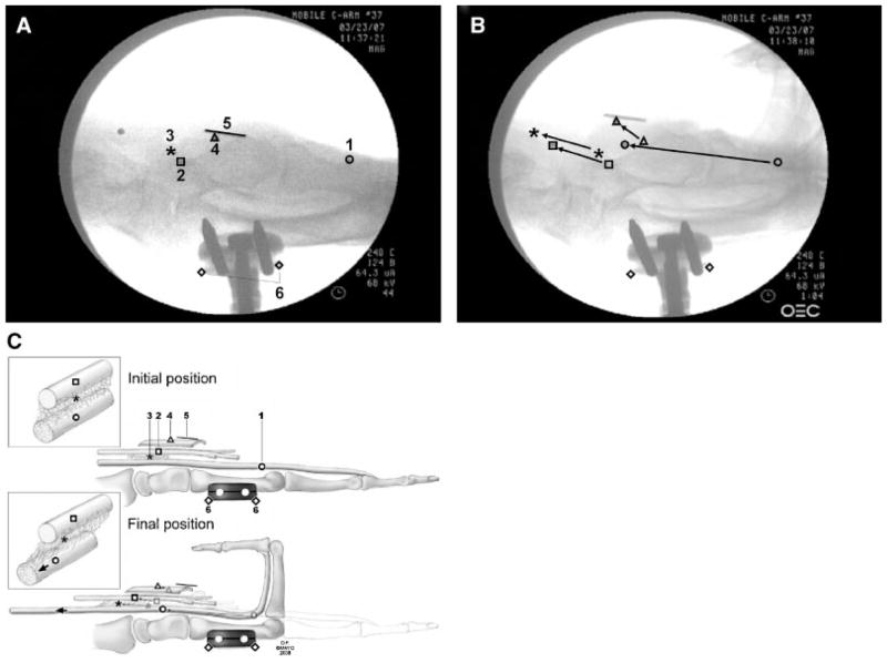
Figure 2.
Example of video frames used to measure marker displacement: (A) initial position and (B) final position. (C) Schematic drawing of initial–final position: 1. tendon; 2. median nerve; 3. SSCT; 4. flexor retinaculum; 5. ruler (10 mm); and 6. the reference points. Copyright© Mayo Foundation; used with permission.

