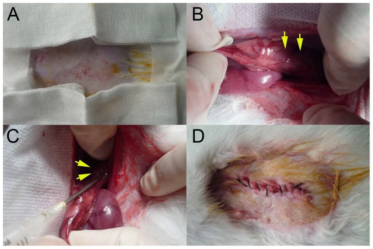
Figure 2. Procedure for surgical implantation.

(A) Hair removal for the operation. (B)The lower thoracic esophagus (yellow arrows) was pulled into the abdominal cavity. (C) Using a 20 G needle, VX2 fragments were inoculated (yellow arrows) into the muscular layer of the esophagus. (D) The abdominal wall was closed.
