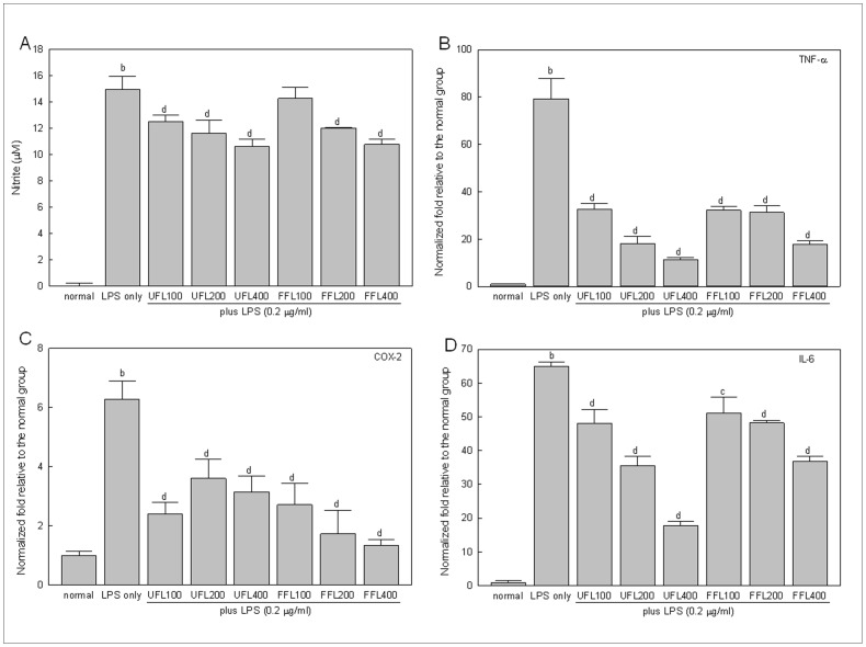
Figure 1. The in vitro anti-inflammatory activities of UFL and FFL in terms of their ability to suppress the LPS-induced production of NO and proinflammatory cytokines.
(A) Immediately after pretreatment with UFL or FFL at 0 (normal, received DMEM instead of herbal extraxt), 100, 200, 400 µg/ml doses for 4 h, RAW 264.7 cells were exposed to DMEM (normal and LPS-alone) or 0.2 µg/ml LPS for 24 h following which NO measurement was performed as described in Materials and Methods section. b P<0.01 compared to the normal and c P<0.05, d P<0.01 compared to the LPS alone. (B–D) Immediately after pretreatment with UFL or FFL at 0 (normal, received DMEM instead of herbal extraxt), 100, 200, 400 µg/ml doses for 4 h, RAW 264.7 cells were exposed to DMEM (normal and LPS-alone) or 0.2 µg/ml LPS for 24 h following which the gene expression profile of proinflammatory cytokines (TNF-α, COX-2 and IL-6) were determined as described in Materials and Methods section. The results are expressed as normalized fold values relative to the normal. b P<0.01 compared to the normal and c P<0.05, d P<0.01 compared to the LPS alone.

