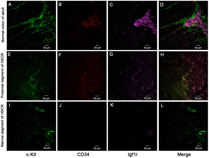
Figure 1. Mature and progenitor ICC located in human adult normal colon by laser confocal.
(A–D) In AP of normal adult colon, the c-Kit positive mature ICCs connected to each other formed extending chords and around AP. ICC progenitors were co-localization of c-Kit+CD34+Igf1r+ found by overlapping fluorescence images. (E–H) In the proximal segment of HSCR, c-Kit positive cells and some of c-Kit+/CD34+/Igf1r+ cells were found occasionally. (I–L) In the narrow segment of the HSCR colon, it was very difficult to find positive ICC and their network structure was damaged, the c-Kit+/CD34+/Igf1r+ cells could not be located by overlapping fluorescence images. Green, red and pink fluorescence represent c-Kit, CD34 and Igf1r, respectively.

