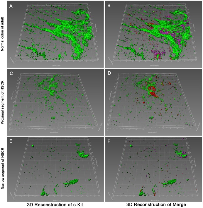
Figure 2. Immunofluorescent illustrations and 3D reconstruction in narrow and proximal segments of HSCR colon, compared with normal adult colon.
(A, B) The 3D network structures of c-Kit+ and c-Kit+/CD34+/Igf1r+ cells were clearly visible in AP of normal adult colon. (C, D) The 3D structures in proximal segment of HSCR were not well formed. (E, F) The 3D structures in narrow segment of HSCR were completely destroyed.

