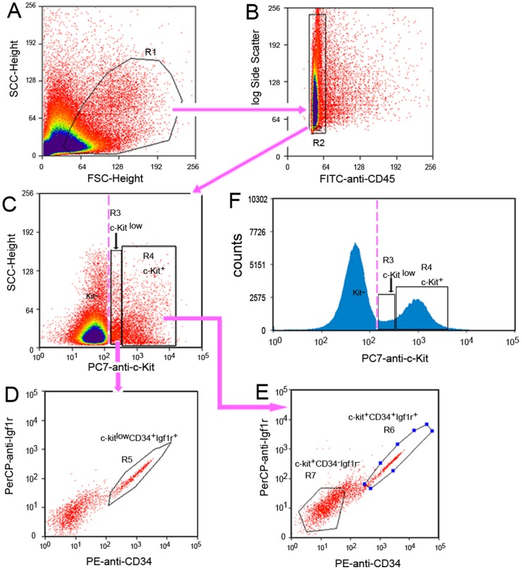
Figure 3. Flow cytometry sorting pathway to purify the mature, early and committed progenitors of ICC.
Analysis following gating: (A) Selection of living mononuclear cells on the histogram with Side Scatter (SSC)/Forward Scatter (FSC). R1 gate was used to select the cells with light scatter properties characteristic of live cells. (B) R2 gate was used to select the cells not expressing macrophage markers (F4/80, CD11b) and the general hematopoietic marker CD45 (FITC- cells): gating on histogram SSC/CD45. (C) Detection of ICC phenotypes: the c-Kit+ cells population was gated in R4 and further analyzed in step E; the c-Kitlow cells population was gated in R3 and further analyzed in next step D. (D) The cells in R5 were c-KitlowCD34+Igf1r+. (E) The cells in R6 were c-Kit+CD34+Igf1r+, and the cells in R7 were c-Kit+CD34−Igf1r−. (F) To confirm the presence of the c-Kitlow population (R3) reliably between c-Kit− and c-Kit+ populations (R4). Pink discontinuous straight line was used as the dividing line between c-Kit− and c-Kitlow populations.

