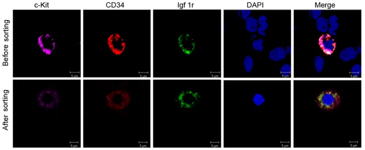
Figure 4. Morphology of single normal human ICC progenitor before and after sorting in suspension.

Upper lane: before sorting, laser confocal microscope displayed phenotypes of ICC progenitor in total cell population from adult normal colon. ICC progenitors showed fluorescence of c-Kit (pink), CD34 (red) and Igf1r (green), merged with blue nuclear stained DAPI. At the same time, other cells only show DAPI staining as negative control, without other fluorescence. Lower lane: after sorting, laser confocal microscopy detected the single intact ICC progenitor with three-color fluorescence. But after immunofluorescence-activated sorting, the fluorescence of ICC progenitors recessed.
