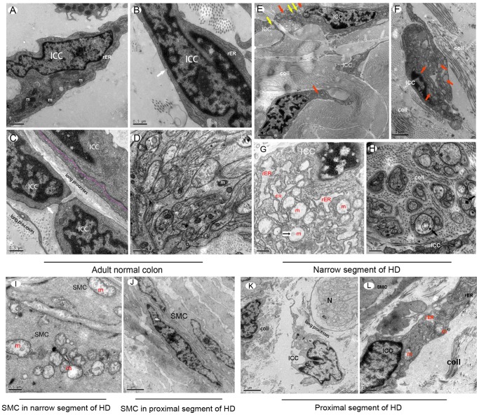
Figure 6. Ultrastructural changes in the narrow and proximal segment of the HSCR colon compared with normal adult colon.
In normal human adult colon, typical ultrastructural features were observed by TEM (A–D). (A) Normal ICC had an oval nucleus with condensed heterochromatin distributed in the periphery, abundant mitochondria (m) and rough endoplasmic reticulum (rER). (B) Caveolae was located along the cell membrane (white arrow). (C) ICCs had long processes. Occasionally they were engaged in multicontact synapses with other ICC (pink dotted line). (D) In AP of human normal colon, there were normal mitochondria (m) inside of normal nerve bundles. (E–G) In the narrow segment of the HSCR colon, ICC showed evidence of severe injury. In almost all of the ICCs, the main features included swollen or vacuolated mitochondria, lack of mitochondrial cristae, dilatated and vesiculated rough endoplasmic reticulum (rER), and degranulated ribosomes. The caveolae on the ICCs plasma membrane were disappeared. Orange arrows in (E) and (F) indicate vesiculated rER. Yellow arrows in (E) indicate swollen or vacuolated mitochondria. (E, F) Injured ICCs were surrounded by large amounts of collagen fibrils (coll), ICC processes were hindered by these collagen fibrils from extending to form connections between cells. (G) High magnification displayed swollen or vacuolated mitochondria and vesiculated rER inside of ICC, which appeared as high density clumps inside of mitochondria (black small arrow). (H) Injured nerve bundles were surrounded by many collagen fibrils (coll). Inside of nerve bundles, swollen mitochondria and lysosomes were identified (black arrows). (I) Swollen or vacuolated mitochondria and dilatated rER inside of smooth muscle cells (SMC) in the narrow segment of the HSCR colon were identified as high density clumps present inside of some SMC mitochondria, which were similar to ICC. (J) Smooth muscle cells in the proximal segment of the HSCR colon were relatively normal. (K, L) In the proximal segment of the HSCR colon, there were long processes of ICC surrounding nerve bundles, not hindered by collagen fibrils (K). There were some swollen mitochondria and dilatated rER inside of ICC (orange font), at the same time some ICC presented with normal rER (black font). A few collagen fibrils (coll) presented beside ICC, but the amount was not greater than in the narrow segment of the HSCR colon.

