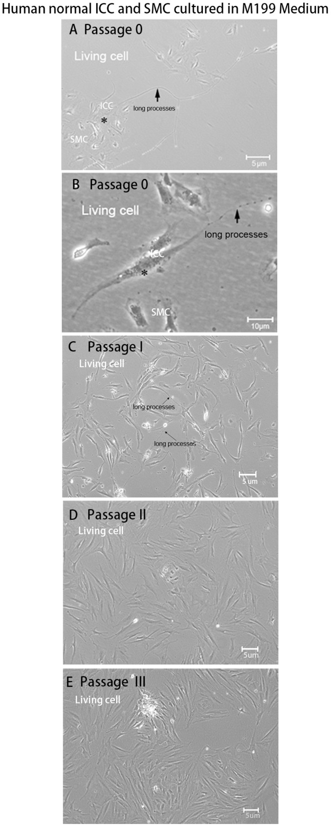
Figure 7. Morphological features of human normal ICC and smooth muscle cells cultured together.

The total cells from adult normal colon muscularis including smooth muscle cells and ICC populations were cultured in M199 without SCF and IGF-I. These cells survived about 20 days. (A, B) Passage 0, the classic descriptions of ICC consistent with previous literature could be observed clearly under a light microscope. The ICC were stellate (A) or spindle (B) shaped, had a different number of ramified long cell processes, and tended to be in contact with smooth muscle cells in culture. (C) Passage 1, ICC with long processes presented in culture. (D, E) With increasing passage number, the primary cells gradually aged so that ICC could not be distinguished from muscle cells and fibroblasts.
