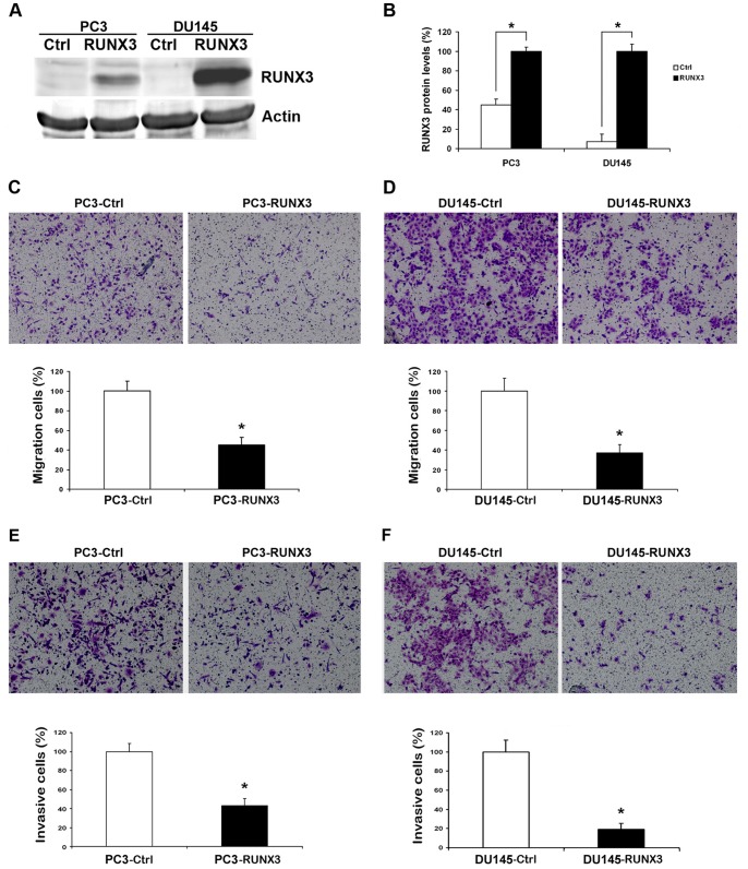
Figure 2. Reduction of RUNX3 on the abilities of metastasis in vitro.
A and B Twenty-four hours after transfection, the expression of RUNX3 in PC3 and DU145 cells was evaluated by Western blot. β-actin was used as an internal control. C and D Cell migration assay. Representative fields of migration cells on the membrane (magnifications, ×200). Average migration cell number per field. E and F Matrigel cell invasion assays. Representative images show the cells that invaded through the Matrigel when transfected with RUNX3 plasmid or control. Representative histograph of invaded tumor cells is displayed and number of invaded tumor cells quantified. * indicates significant difference from the controls (*P<0.05, ANOVA).

