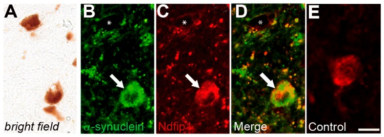
Figure 4. Ndfip1 is expressed in dopaminergic neurons containing α-synuclein deposits.

(A) Bright field image of dopaminergic neurons in the substantia nigra of a PD brain. (B–D) Fluorescent labelling of α-synuclein and Ndfip1 from the bright field image shows deposits of α-synuclein which co-label with Ndfip1 in a dopaminergic neuron (arrow). Asterisk marks a dopaminergic neuron with neither α-synuclein nor Ndfip1 positive labelling. (E) Fluorescent labelling of Ndfip1 in a control brain. Scale bar: 25 µm.
