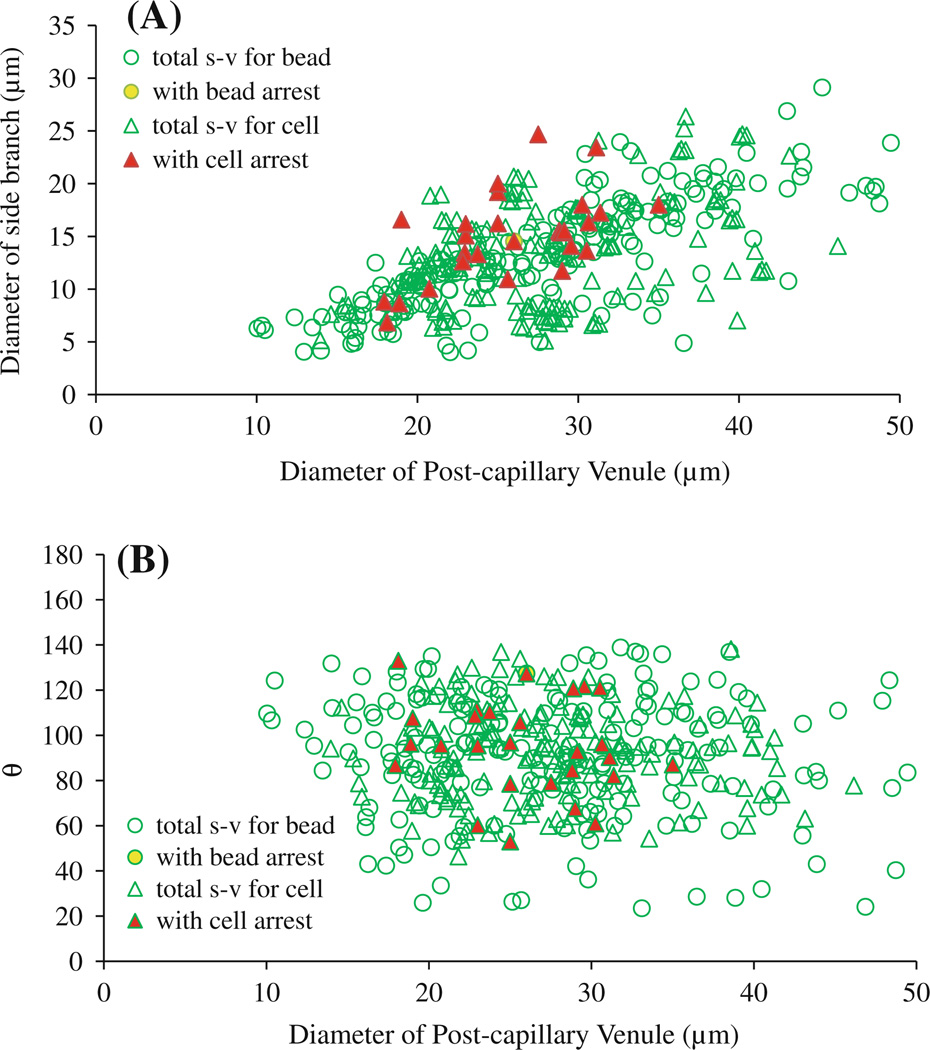
Fig. 5.
Diameters and branching angles of microvessels at capillary (or postcapillary venule)–postcapillary venule intersections with potential arrest of cells/beads. A Diameter of postcapillary venules versus diameter of side branches at capillary (or postcapillary venule)–postcapillary venule intersections.B Diameter of postcapillary venules versus branching angle θ between postcapillary venule and side branch. Empty green circles: total side branch–postcapillary venule (s–v) intersections examined in five rats injected with beads; yellow filled circles: intersections with arrested beads. Empty green triangles: total side branch–postcapillary venule intersections examined in six rats injected with tumor cells; red filled triangles: intersections with arrested cells

