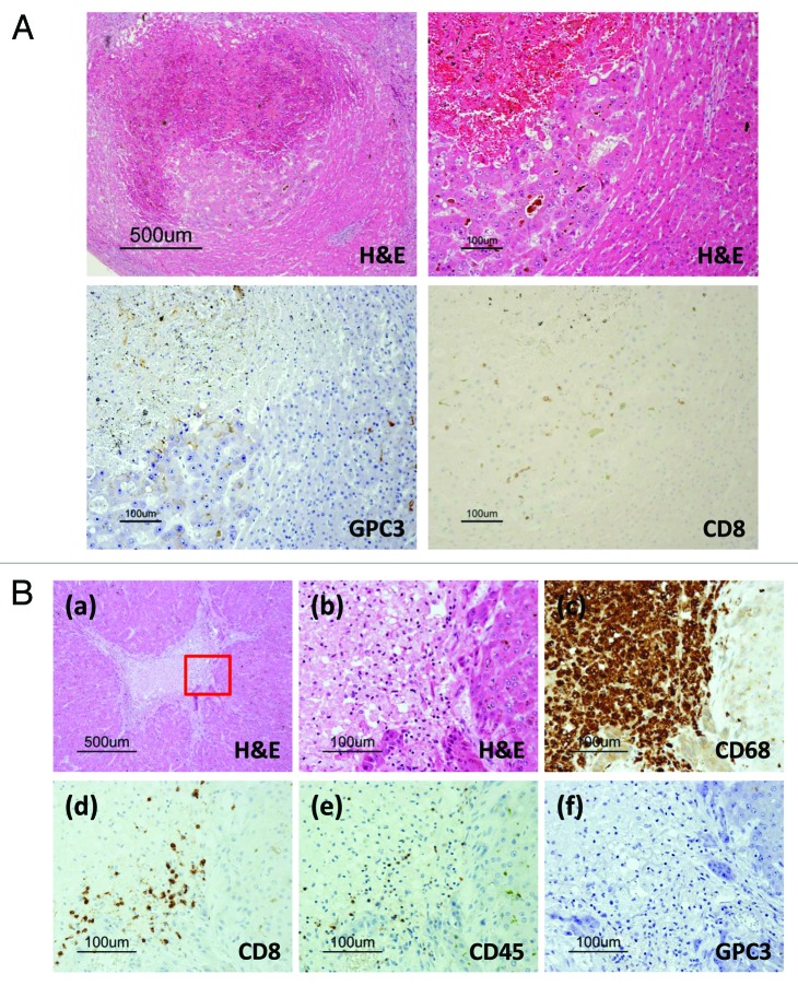
Figure 3. Pathological findings in the autopsy specimen. (A) Carcinoma in a cirrhotic nodule. CD8-positive T cells (brown) infiltrated only the carcinoma area, accompanied by necrosis. No infiltration of CD8-positive T cells was detected in the cirrhotic nodule. Only carcinoma cells were GPC3-positive by immunohistochemical staining. (B) Necrotic area surrounded by cirrhotic nodules. (a) Necrosis was surrounded by viable cirrhotic cells. (b) The margin between the necrosis and the cirrhotic nodule. This portion is enclosed by the red box in (a). (c) CD68-positive macrophages (brown) aggregated in the necrotic area around the cirrhotic nodule. (d) CD8-positive T cells (brown) infiltrated the necrotic area but not the cirrhotic nodule. (e) CD45-positive lymphocytes infiltrated the necrotic area. Based on the image in (d), most of the lymphocytes were CD8-positive T cells. (f) Cirrhotic cells did not express GPC3.

An official website of the United States government
Here's how you know
Official websites use .gov
A
.gov website belongs to an official
government organization in the United States.
Secure .gov websites use HTTPS
A lock (
) or https:// means you've safely
connected to the .gov website. Share sensitive
information only on official, secure websites.
