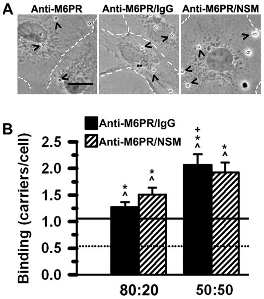
Figure 3. Effect of surface functionalization with NSM on the binding of anti-M6PR carriers.
(A) Phase-contrast microscopy showing binding of 1-μm parent anti-M6PR carriers compared to 50:50 anti-M6PR/IgG carriers and anti-M6PR/NSM carriers, 3 h after incubation at room temperature with fixed activated HUVECs (dashed lines mark cell borders). Scale bar = 10 μm. (B) Binding of anti-M6PR/IgG carriers vs. anti-M6PR/NSM carriers (for both 80:20 and 50:50 formulations) was determined by phase-contrast microcopy as in Fig. 1A. Mean ± standard error of the mean. *Compared to parent anti-M6PR carriers (continuous line in graph); ^compared to control IgG carriers (dotted line in graph); +comparison between different surface-coverage ratios (80:20 vs. 50:50) of the same formulation type (p < 0.05). No difference was found comparing anti-M6PR/IgG vs. anti-M6PR/NSM carriers.

