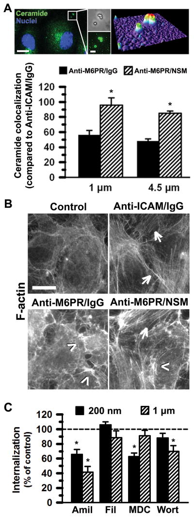
Figure 6. Mechanism associated with NSM-functionalization of carriers.
(A) Enhanced colocalization of 1-μm or 4.5-μm anti-M6PR/NSM carriers (50:50 surface-coverage ratio) with ceramide at the surface of activated HUVECs after 30 min incubation at 37°C, compared to anti-M6PR/IgG carriers. An example of 1-μm anti-M6PR/NSM carriers (phase contrast) colocalizing with ceramide (green fluorescence) and the corresponding fluorescence surface-plot (upper right) is shown. Blue = DAPI-positive cell nuclei. Scale bars = 10 μm (left) and 2 μm (right). Mean ± standard error of the mean. *Compares anti-M6PR/NSM carriers to anti-M6PR/IgG counterparts (p < 0.05). (B) F-actin staining with fluorescent phalloidin shows stress fibers in activated HUVECs after incubation with control media or media containing 1-μm anti-M6PR/NSM carriers (50:50) for 30 min at 37°C. Anti-M6PR/IgG carriers are shown as negative controls, while anti-ICAM/IgG carriers are positive controls. Arrows indicate stress fibers and arrowheads indicate ruffling. Scale bar = 10 μm. (C) Percent endocytosis of 200-nm and 1-μm anti-M6PR/NSM carriers (50:50) was assessed using activated HUVECs incubated for 3 h at 37°C in the absence (control) vs. presence of pharmacological inhibitors of pathways mediated by clathrin (monodalsylcadaverine, MDC), caveolae (filipin, Fil), CAM and macropinocytosis (amiloride, Amil), or macropinocytosis (wortmannin, Wort). Mean ± standard error of the mean. *Compares inhibitor to control for each carrier size (p < 0.05).

