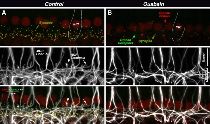
Fig. 3.
Ouabain can eliminate virtually all synapses between auditory nerve fibers (ANFs) and inner hair cells (IHCs), as seen in maximal projections of confocal z-stacks. In the normal ear (A), each synapse (top panel) is a juxtaposed pair of red (anti-CtBP2) and green (anti-GluA2) puncta, showing the presynaptic ribbon and the postsynaptic receptor patch, respectively. IHC nuclei are also faintly stained (red), and the rough outline of one IHC is shown (dotted line). Unmyelinated processes of ANFs and medial olivocochlear fibers (MOC) are stained with anti-neurofilament antibodies (middle panel). MOC fibers project to outer hair cells outside the field of view. In the merge view (bottom panel), the juxtaposition between ANF terminals and synaptic puncta is evident (see white arrows). In the ouabain ear (B), only one synapse remains (red-green arrow) among these ten IHCs. There are numerous orphan ribbons (e.g., red arrow) and two orphan receptor patches (e.g., green arrow). The merged view (bottom panel) suggests that these orphan elements are not paired with ANF terminals (e.g., arrows). Images are from the 32 kHz region; scale bar in A (merge) applies to all panels.

