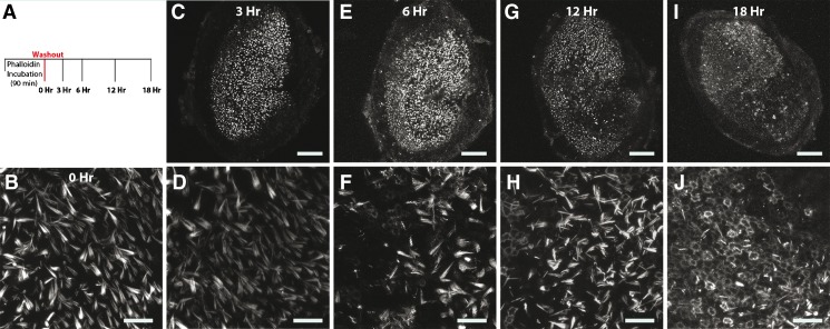
FIG. 2.
Time series of fluorescence observed after culturing utricles that had been treated with phalloidin-CF488A. Fluorescence disappears from hair cells and appears within circumferential F-actin belts of supporting cells in culture. A Experimental outline. Utricles were treated with phalloidin-CF488A for 90 min, then the label was washed out three times with DMEM/F-12, and then the media was replaced with DMEM/F-12 containing 1 % FBS. Utricles were then maintained in culture at 37 °C. Utricles were mounted and imaged live immediately (0 h; B), or after 3 h (C, D), 6 h (E, F), 12 h (G, H), or 18 h in culture (I, J). Around 6 h, the label begins to disappear from hair cells and supporting cells become fluorescent (F), and by 18 h, nearly all supporting cells are fluorescent (J). Scale bars in B, C, E, G, and I are 100 μm; scale bars in D, F, H, and J are 20 μm.

