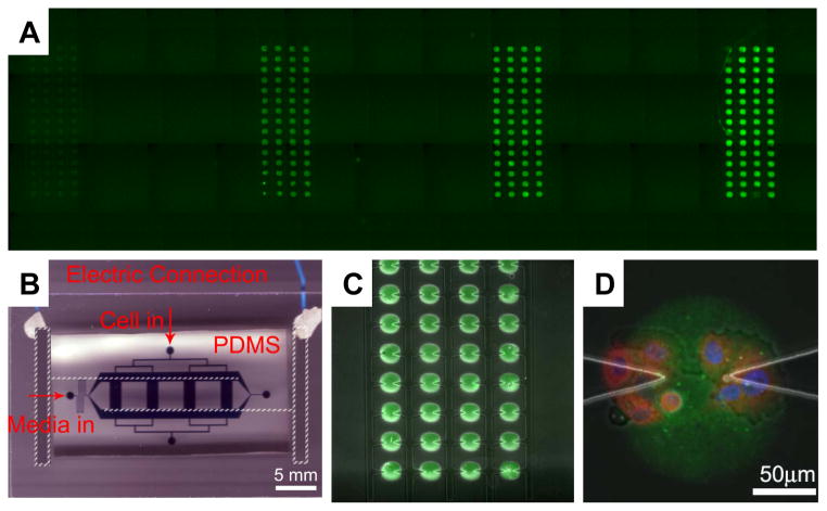
Figure 2.
Combining dielectrophoresis-assisted microfluidics with protein micro-arraying for cell signaling studies. A: Four columns of Alexa 488 conjugated goat-anti-mouse antibody printed onto the glass substratum part of the device, with concentration increasing from left to right, 0.001, 0.01, 0.1 and 1 mg/ml, respectively; B: A photograph of a fully assembled functional device, highlighting the electrical wiring access to the on-glass ITO electrodes (parts not readily visible indicated in white broken lines), the PDMS microfluidic chips with channels filled with a food dye and inlets - annotated; C: Demonstration of the alignment of a part of the in-chip protein array (green spots indicating the printed antibody as in (A)) with the triangular electrode array inside one of the fluidic channels; D: One Oregon Green 488 conjugated collagen IV array spot (25 μg/ml coating concentration) with several iHUVECs captured and cultured on it. Cells were stained within the chip to indicate the location of the nucleus (stained with DAPI, blue) and anti-p65 antibody (red).

