
Figure 2. Computed Tomographic Scans of Patients with Graves’ Ophthalmopathy and of a Normal Subject.
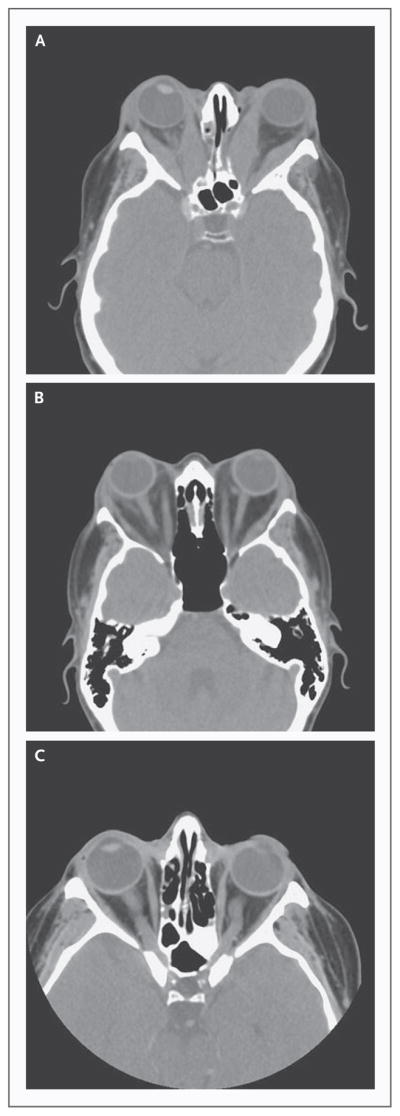
Axial images of patients with Graves’ ophthalmopathy reveal generalized enlargement of the extraocular muscles with marked bilateral proptosis (Panel A) and marked bilateral proptosis and asymmetric involvement of the extraocular muscles with expansion of the orbital fat bilaterally (Panel B). Normal orbits are shown (Panel C) for comparison.