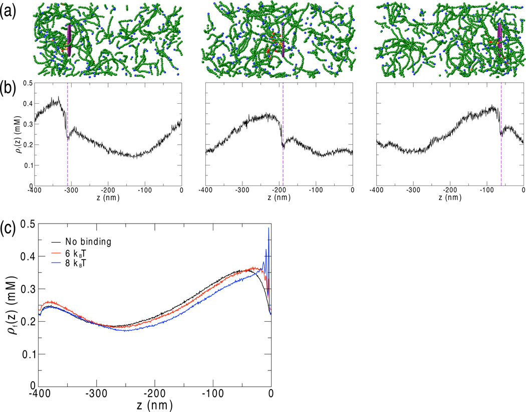
Figure 1.
(a) F-actin (green) accumulates behind the disk (purple). Pointed ends are shown in blue, bases of actin branches are shown in red, and subunits attached to the disk are shown in orange. For clarity, G-actin is not displayed. The system shown has 50 6 kBT binding sites randomly distributed on the back of the disk (ρActA = 0.64 per 100 nm2). Axes and scale match those of panel (b), below. (b) The F-actin concentration profile along the z-direction is potted at times corresponding to the snapshots shown above in (a). As described in [13], Arp2/3-mediated F-actin polymerization generates a concentration gradient of F-actin behind the disk. As the disk moves forward, F-actin continues to polymerize behind the disk so that the concentration gradient moves forward as well, maintaining the mechanism of motility. (c) In the center of mass frame of the disk, which is located at z = 0, the concentration gradient has a well-defined steady-state average that depends on the binding energy.

