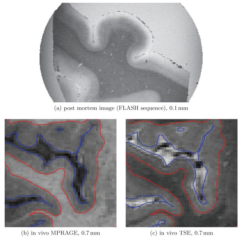
Figure 1.
Post mortem tissue scanned at 0.1 mm with a FLASH sequence (a) and the two contrasts we acquired for our study (b-c). Each image is in coronal orientation and centered to Heschl’s gyrus. In (a) the lower two thirds of the gray matter in this region clearly show a shift in intensity similar to the white matter. This, however, is not apparent in the in vivo images. Also note that the lower layers seem to be compressed within deep sulci left and right of Heschl’s gyrus in Figure (a).

