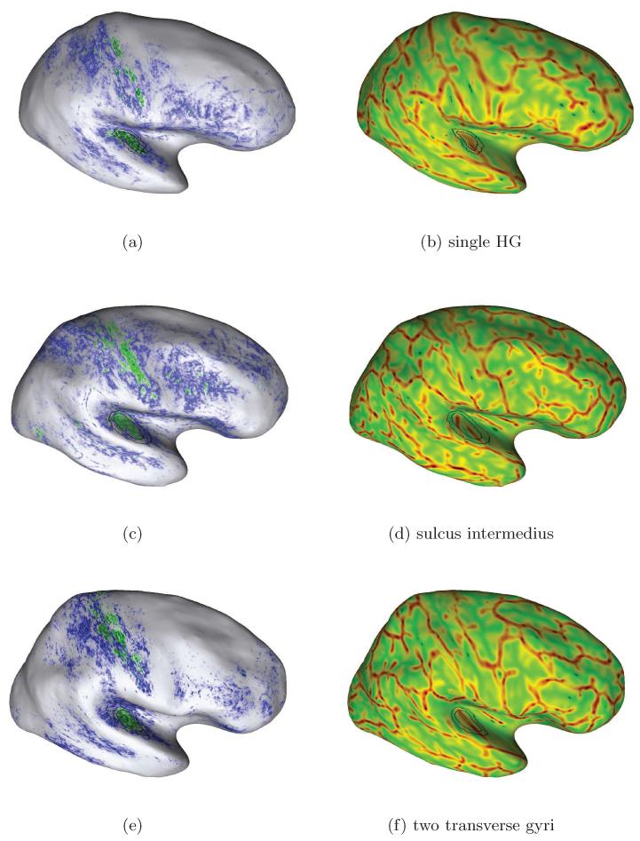
Figure 8.
Additional examples of individual mapping results due to our approach for in-vivo localization of the human PAC area. Here, we selected further subjects with different temporal cortex anatomy in the right hemispheres: Heschl’s gyrus with (second row) and without sulcus intermedius (top and bottom) and one subject with an additional transverse temporal gyrus (bottom row). A higher myelinated region (green labelling) of plausible size can be identified on the medial two thirds of Heschl’s gyrus in each case (cf. Sect. 3.2).

