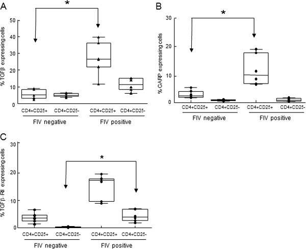
Figure 1.

PBMCs from FIV-infected cats display increased surface expression of TGFβ and GARP on CD4+CD25+lymphocytes and TGFβRII on CD4+CD25-lymphocytes. PBMCs from FIV-infected or control cats were analyzed for surface expression of CD4, CD25, TGFβ, GARP and TGFβRII by flow cytometry. Cells were gated on CD4+CD25+ or CD4+CD25- populations and analyzed for percent expression of A. TGFβ, B. GARP or C. TGFβRII. Box-whisker plots represent the 5th and 95th percentiles (whisker), 25th and 75th percentiles (box), and median of percent CD4+CD25+ and CD4+CD25- expression from 6 FIV-positive and 6 FIV-negative cats. Symbols represent individual cats. CD4+CD25+ cells from FIV+ cats exhibit greater surface expression of TGFβ and GARP when compared to CD4+CD25+ cells from FIV- cats. CD4+CD25- cells from FIV+ cats exhibit greater surface expression of TGFβRII than CD4+CD25- cells from FIV- cats. (p <0.05, Mann–Whitney test for significance).
